The cost to develop and launch a new drug in the market can be, by some estimates, as staggeringly high as $4.54 Bn. It is also remarkably tedious, sometimes taking up to 15 years to make a drug commercially available. Nowadays, clinicians often employ the computer-assisted Drug Design or CADD technique to speed up the process.
Computational intelligence can be utilized to perform an array of drug discovery analyses. Artificial techniques, such as automated cell counting methods, not only help scientists determine the molecular properties of the end product. They can also make intelligent recommendations about the types of compounds that will help achieve the desired drug.
Table of Contents
Barriers to Drug Discovery & Development
Scientists and doctors continuously try to discover and develop new drugs to treat diseases such as neurodegenerative disorders, cancer, etc. Unfortunately, drug development is a tedious and expensive process, vulnerable to failure.
A drug that can be successfully marketed can cost more than $1 Bn and up to 15 years to develop. The pre-clinical stage, which can take up to 6 years, uses animal models to test the efficacy and safety of the drug. It also identifies, validates, and optimizes human candidates for drug trials.
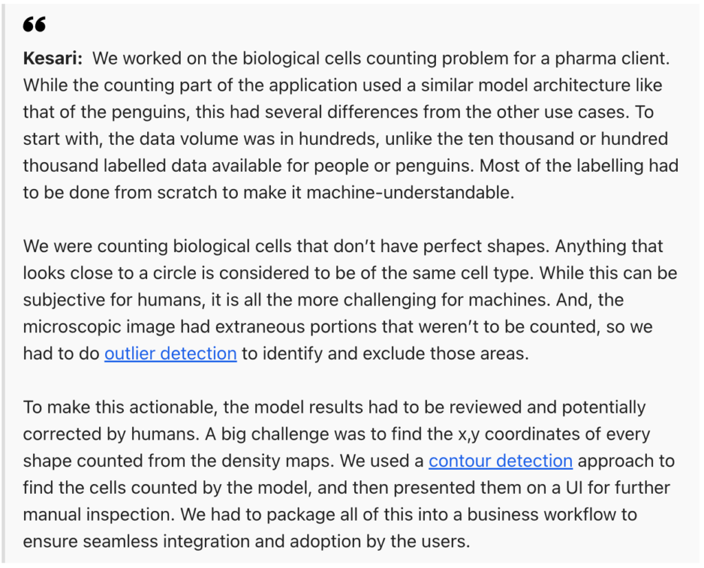
“Drug characterization is a key step in the pharma drug discovery process. Scientists need counts of different cell types from microscopic images. This painful, manual process is being automated by the algorithms.”
Quote for InfoQ by Ganes Kesari – Chief Decision Scientist, Gramener
In the US, clinical trials can only begin after the FDA accepts the Investigational New Drug (IND) application.
In the first clinical phase, one-third of the drugs fail. Despite the animal models predicting the drugs to be non-toxic to humans, about half of the human participants in the clinical trials succumb to unforeseen toxicity at some point.
Furthermore, around a quarter of the clinical trial drugs have no effect on humans.
The Importance of Cell Analysis in Drug Development
Healthy and diseased tissues differ in composition, function, and genetics. This poses a challenge for drug development and discovery. It is a barrier to developing accurate disease models that can be used to design drugs.
It can also make it a challenge to gauge the responses of patients to specific therapies.
Studying heterogeneous tissues and their complex nature has led to the development of multiple cell analysis tools.
Due to the differences in cell populations within biological systems, individual cell analyses are crucial to identifying mechanisms that cannot be studied in groups of cells.
Cell populations, even within the same species, can significantly vary in terms of their physiological and biological functions. Individual cell analyses can help scientists understand the underlying factors that cause diseases.
The ability to analyze at single-cell and multi-cell levels enable drug developers to gain critical insights. It gives researchers an all-encompassing and balanced picture.
Statistical analyses of entire cell populations offer aggregated insights and are non-representative of any single cell, especially rare variants. Analyzing individual cells within a heterogenous population reduces biological noise.
It also helps scientists characterize and investigate rare cells.
Challenges to Traditional Cell Analysis
One initial challenge of single-cell analysis is the isolation and culture of an individual cell. Cell viability is required for further cell outgrowth and downstream analysis.
Cells are heavily influenced by their interactions with other cells and their surrounding environment. They do not exist individually in nature and can be hard to culture separately.
Furthermore, individual cells removed from their natural surroundings may respond unpredictably to an artificial environment. The isolation process may change the profile, viability, and behavior of the cell and can even result in cell stress.
Processing power is another challenge in cell analysis. To analyze each cell individually, millions of single cells must be processed in a fast and accurate fashion.
Unfortunately, many analysis techniques destroy the cells in the process. Additionally, it can be challenging to separate biological noise from the relevant information when analyzing a heterogeneous cell population.
Advances in the platforms and methods to analyze individual cells, such as automated cell counting techniques, have vastly increased the amount of data produced. A single cell may produce detailed data in vast arrays of information, requiring specialized interpretation and analysis.
Application of AI in Drug Discovery
Protein dysfunctions lead to impurities in the human body. Drug design strategies involve the differentiation of small molecules within protein targets. It involves a substantial amount of time and money to predict the 3D structure of proteins, with poor accuracy rates.
Deep learning and feature extraction tools can help gain crucial information on the connection between protein sequence and structure. In the future, it can even improve the accuracy of predicting the 3D protein structure.
Artificial Intelligence is used in applications like ensuring the safety of biomarkers, the discovery of drug efficacy, drug-protein interaction prediction, and automated cell counting.
Computational techniques can predict the protein targets for distributed drug molecules. Network-based systems can help with drug repurposing, conflict detection, and predicting associations for novel drug diseases.
Deep learning techniques can also ensure the safety of biomarkers.
Read more: The Role of Artificial Intelligence in Drug Discovery and Pharmaceutical Research
Gramener Offers Never-before Advantages in Early Drug Discovery
At Gramener, we specialize in helping pharmaceutical companies reduce their overall efforts using computer vision applications, such as automated cell counting software. We enabled one of our top pharma clients to reduce their manual effort by up to 95% and increase accuracy by more than 90%.

Counting cells in liposome images using a Transmission Electron Microscope led to high manpower requirement, high-cost consumption, and up to 35% manual error.
Using a Convoluted Neural Network (CNN) based Deep Learning counting model, we could detect and count the number of cells in a liposome image. An interactive UI improved the interaction between humans and computers.
Contact us for custom built low code data and AI solutions for your business challenges and check out pharma and life sciences AI solutions built for our clients, including Fortune 500 companies. Book a free demo right now.

